For our international customers, please be advised that orders cannot be placed through our website by customers in countries with International Distributor representation.
Dextranase - Manual
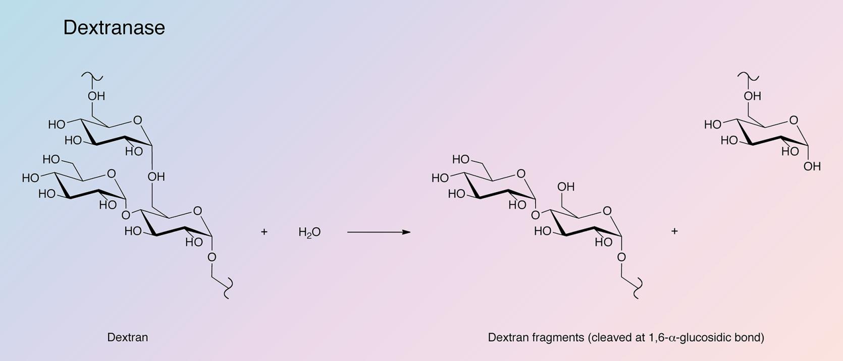
Dextranases catalyze the endohydrolysis of 1,6-alpha-glucosidic linkages in dextran.
The search for organisms producing large amounts of an enzyme to break down dextran began in the 19th century (van Tieghem 1878).Organisms producing trace amount of both intracellular and extracellular dextranase were isolated in the late 1940s (Ingelman 1948, and Hultin and Nordström 1949) but it was not until the 1950s that a Penicillium species producing large amounts of extracellular dextranase was reported (Tsuchiya et al. 1952).
In the early 1970s, the first purifications and characterizations of Penicillium dextranase were being conducted (Baastad and Rolla 1970, Chaiet et al. 1970, and Fukumoto et al. 1971). In the later 1970s, research began to explore the use of dextranase for the treatment of tooth decay (Minah et al. 1972, Goldstein-Lifschitz and Bauer 1976, and Simonson et al. 1979).
In the 1990s, the Penicillium dextranase gene was cloned and expressed in Pichia pastoris (Garcia et al. 1996, and Roca et al. 1996). An early classification system for dextranase and other dextran-hydrolyzing enzymes was also developed using sequence-analysis software (Aoki and Sakano 1997).
The crystal structure of Penicillium minioluteum was elucidated by Larsson et al. in 2003. This study also importantly suggested the reaction mechanism proceeds by net inversion (rather than retention) of the anomeric carbon. Recent research has aimed to improve enzyme stability of fungal dextranase using site-directed mutagenesis (Chen et al. 2009).
Specificity:
Dextranase preferentially cleaves the alpha-1,6 linkages of dextran, releasing shorter isomaltosaccharides, with net inversion of the anomeric carbon configuration (Larsson et al. 2003, and Sugiura et al. 1973).
Molecular Characteristics:
Extracellular dextranase is encoded by the dex gene. Post-translational modification includes cleavage of the signal peptide and N-glycosylation. Amino acid sequence analyses have revealed 29% identity between Penicillium minioluteum and Arthrobacter sp. (Roca et al. 1996).
Composition:
Dextranase enzymes belong to two glycoside hydrolase families, either 49 or 66, which do not share significant sequence similarity. Dextranases from Penicillium and Arthrobacter species are classified as family 49, while those of the Streptococcus species are found in family 66 (Coutinho and Henrissat 1999).
Penicillium dextranase and related enzymes contain two domains. The first domain resembles the immunoglobulin fold and consists of 200 amino acid residues forming 13 beta strands. A beta sandwich is formed by nine of those strands, with all but strands 5 and 13 being antiparallel. The second domain contains a right-handed parallel beta-helical fold containing 3 parallel beta sheets (Larsson et al. 2003). These two domains are connected by a large interface, which contains 29 amino acids completely conserved in the glycoside hydrolase family 49. Disulfide bridges are formed by four of the six cysteines present in the protein, none of which are conserved within the glycoside hydrolase family 49 (Larsson et al. 2003).
Protein Accession Number: CAB91097
CATH Classification:
- Class: Mainly Beta
- Architecture: Sandwich; 3 Solenoid
- Topology: Dex49a from Penicillium minioluteum complex, domain 1; Pectate Lyase C-like
Molecular Weight:
- 64.6 kDa (Theoretical)
Optimal pH Range:
- 5.0-7.0 (Chaiet et al. 1970)
Isoelectric Point:
- 4.55 ± 0.05 (Chaiet et al. 1970)
- 3.88 (Raices et al. 1991)
Extinction Coefficient:
- 119,350 cm-1M-1 (Theoretical)
- E1%,280 = 20.0 (Chaiet et al. 1970)
Activators:
- Co2+, Mn2+, Cu2+ (Sugiura et al. 1973)
Inhibitors:
- Hg2+, Ag2+
- N-bromosuccinimide
- I2
(Sugiura et al. 1973)
Applications:
- Viscosity reduction in cell culture (Lindskog et al. 1987)
- Glycobiology
- Reduction in viscosity caused by dextran during sugar processing (Larsson et al. 2003)
